


eBook
Available on Compatible NOOK devices, the free NOOK App and in My Digital Library.
Related collections and offers
Overview
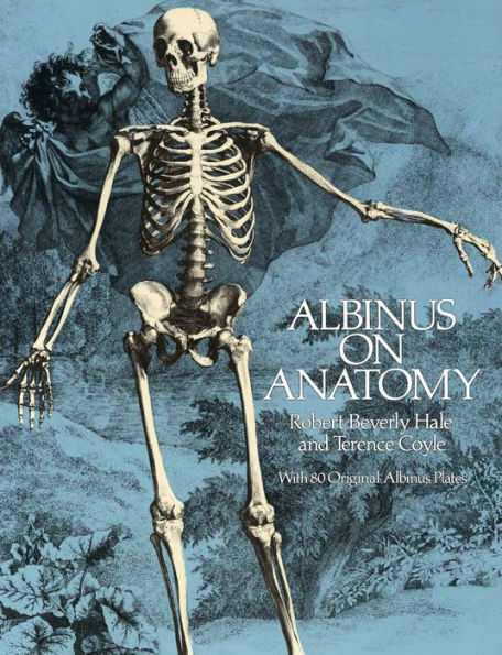
All 80 of the original copperplate engravings, containing over 230 individual illustrations, have been painstakingly reproduced for this edition. The muscles and bones of the human body are rendered individually and in related groups from varying perspectives, enabling art students to compare the forms; to analyze their size, shape, direction, and attachments; and to observe with absolute clarity the shape and position of bodily forms. Eighty modern diagrams matching each plate identify each bone and muscle in the most common medical terms.
The eminent medical historian Charles Singer praised Albinus' brilliant accomplishment: "He introduced a new standard of accuracy into practical anatomy and of accuracy and beauty into anatomical illustrations." Singer adds: "These illustrations, with their finely wrought ornamental backgrounds, were intended for artists as well as for physicians, and no finer work of their type has ever been executed."
Introductory essays by the well-known artist and art educator Terence Coyle — including a new introduction to the Dover edition — engagingly explore Albinus' life and work. Following these, Robert Beverly Hale, one of America's best-known teachers of figure drawing and anatomy, brilliantly appraises Albinus' technique and demonstrates how artists today can use his anatomical studies to draw from life, a special feature that makes this magnificent book truly indispensable for artists and art students at every level.

Product Details
| ISBN-13: | 9780486318868 |
|---|---|
| Publisher: | Dover Publications |
| Publication date: | 04/29/2013 |
| Series: | Dover Anatomy for Artists |
| Sold by: | Barnes & Noble |
| Format: | eBook |
| Pages: | 208 |
| File size: | 28 MB |
| Note: | This product may take a few minutes to download. |
Read an Excerpt
ALBINUS ON ANATOMY
With 80 Original Albinus Plates
By Robert Beverly Hale, Terence Coyle
Dover Publications, Inc.
Copyright © 1979 Watson-Guptill PublicationsAll rights reserved.
ISBN: 978-0-486-31886-8
CHAPTER 1
THE BONES AND MUSCLES OF THE BODY
THE SKELETON, FRONT VIEW
1. Frontal bone
2. Superciliary eminence
3. Orbit
4. Nasal bone
5. Superior maxillary (Maxilla, upper jaw)
6. Inferior maxillary (Mandible, lower jaw)
7. Clavicle
8. Acromion process of scapula
9. Coracoid process of scapula
10. Scapula
11. Sternum
12. Humerus
13. Radius
14. Ulna
15. Carpals
16. Metacarpals
17. Phalanges
18. High point of pelvis
19. Iliac tubercle (Wide point)
20. Parietal bone
21. Temporal bone
22. Zygomatic (Malar, cheek bone)
23. Mastoid process of temporal bone
24. Ramus of mandible
25. Cervical vertebrae
26. First rib
27. Fifth rib
28. Thoracic (dorsal) vertebrae
29. Line where rib meets cartilage
30. Tenth rib
31. Lumbar vertebrae
32. Iliac crest
33. Anterior superior iliac spine (Front point)
34. Ilium of pelvis
35. Sacrum
36. Pubis
37. Great trochanter of femur
38. Ischium
39. Lesser trochanter of femur
40. Shaft (body) of femur
41. Patella
42. Outer epicondyle of femur
43. Tibia (Shin bone)
44. Fibula
45. Outer (lateral) malleolus of fibula (Outer ankle)
46. Tarsals
47. Metatarsals
48. Phalanges
49. Calcaneus (Heel bone)
THE SKELETON, BACK VIEW
1. Occipital bone
2. Atlas (First cervical vertebra)
3. Axis (Second cervical vertebra)
4. First rib
5. Clavicle (Collar bone)
6. Acromion process of scapula
7. Humerus
8. Radius
9. Ulna
10. Styloid process of ulna
11. Carpals
12. Metacarpals
13. Phalanges
14. Olecranon of ulna (Elbow)
15. Fifth rib
16. Line of angle of ribs
17. Tip of tenth rib
18. Lumbar vertebrae
19. High point of pelvis
20. Ilium of pelvis
21. Sacrum
22. Great trochanter of femur
23. Coccyx
24. Ischium
25. Lesser trochanter of femur
26. Parietal bone
27. Vertebra prominens (Seventh cervical vertebra)
28. Scapula
29. Dorsal (thoracic) vertebrae
30. Shaft (body) of femur
31. Outer epicondyle of femur
32. Outer condyle of femur
33. Tibia
34. Fibula
35. Outer malleolus of fibula
36. Tarsals
37. Metatarsals
38. Phalanges
39. Inner epicondyle of femur
40. Inner condyle of femur
41. Inner malleolus of tibia
42. Calcaneus (Heel bone)
THE SKELETON, SIDE VIEW
1. Frontal bone
2. Zygomatic (Malar, cheek bone)
3. Superior maxillary (Maxilla, upper jaw)
4. Inferior maxillary (Mandible, lower jaw)
5. Clavicle
6. Spine of scapula
7. Humerus
8. Olecranon of ulna (Elbow)
9. Ulna
10. Radius
11. Carpals
12. Metacarpals
13. Phalanges
14. Fifth rib
15. Line where rib meets cartilage
16. Tip of tenth rib
17. Lumbar vertebrae
18. High point of pelvis
19. Iliac tubercle of pelvis (Wide point)
20. Anterior superior iliac spine (Front point)
21. Ilium of pelvis
22. Anterior inferior iliac spine (Secondary point)
23. Pubis
24. Great trochanter of femur
25. Ischium
26. Shaft (body) of femur
27. Patella (Kneecap)
28. Inner epicondyle of femur
29. Tibia
30. Fibula
31. Parietal bone
32. Occipital bone
33. Atlas (First cervical vertebra)
34. Axis (Second cervical vertebra)
35. Vertebra prominens (Seventh cervical vertebra)
36. First rib
37. Scapula
38. Dorsal (thoracic) vertebrae
39. Inner (medial) epicondyle of humerus
40. Posterior superior iliac spine (Back point)
41. Posterior inferior iliac spine
42. Sacrum
43. Coccyx
44. Outer (lateral) epicondyle of femur
45. Calcaneus (Heel bone)
46. Phalanges
47. Metatarsals
48. Tarsals
THE OUTERMOST ORDER OF MUSCLES, FRONT VIEW
1. Frontalis
2. Nasalis (Compressor naris)
3. Orbicularis oris
4. Pectoralis major
5. Coracobrachialis
6. Biceps brachii, outer (long) head
7. Biceps brachii, inner (short) head
8. Triceps, middle (scapular or long) head
9. Triceps, outer (long humeral) head
10. Triceps, inner (short humeral) head
11. Brachialis
12. Brachioradialis (Supinator longus)
13. Extensor carpi radialis longus
14. Flexor digitorum superficialis (middle layer)
15. Flexor pollicis longus
16. Abductor pollicis brevis
17. Pronator teres
18. Flexor carpi radialis
19. Palmaris longus
20. Flexor carpi ulnaris
21. Palmaris brevis
22. Orbicularis oculi (palpebrarum)
23. Zygomaticus major
24. Masseter
25. Sternocleidomastoid
26. Platysma
27. Trapezius
28. Deltoid, anterior (clavicular) portion
29. Deltoid, middle (acromionial) portion
30. Extensor carpi radialis brevis
31. Extensor digitorium
32. Abductor pollicis longus
33. Extensor pollicis brevis
34. Abductor of index (First dorsal interossei)
35. Latissimus dorsi
36. Serratus anterior
37. External oblique (Obliquus externus)
38. Rectus abdominus
39. Linea alba
40. Umbilicus (Navel)
41. Anterior superior iliac spine (Front point)
42. Inguinal (Poupart's) ligament (Line of groin)
43. Gluteus medius
44. Pyramidalis
45. Iliacus
46. Psoas
47. Tensor fasciae latae
48. Pectineus
49. Sartorius
50. Adductor longus
51. Gracilis
52. Adductor magnus
53. Rectus femoris
54. Vastus externus (lateralis)
55. Vastus internus (medialis)
56. Patella
57. Head of fibula
58. Anterior tuberosity (Kneeling point)
59. Peroneus longus
60. Soleus
61. Gastrocnemius
62. Tibialis anterior
63. Extensor digitorum longus
64. Anterior annular ligament
65. Semitendinosus tendon
66. Sartorius tendon
67. Abductor hallucis
68. Extensor hallucis longus
THE OUTERMOST ORDER OF MUSCLES, BACK VIEW
1. Occipitalis
2. Sternocleidomastoid
3. Deltoid, posterior (scapular) portion
4. Deltoid, middle (acromionial) portion
5. Triceps, middle (scapular or long) head
6. Triceps, outer (long humeral) head
7. Brachialis
8. Brachioradialis (Supinator longus)
9. Extensor carpi radialis longus
10. Extensor carpi radialis brevis
11. Extensor digitorum
12. Extensor digiti minimi
13. Extensor carpi ulnaris
14. Triceps, inner (short humeral) head
15. Palmaris longus
16. Anconeus
17. Flexor digitorum profundus (deep layer)
18. Flexor carpi ulnaris
19. Flexor digitorum superficialis (middle layer)
20. External oblique
21. Iliac crest
22. Gluteus medius
23. Gluteus maximus
24. Tensor fasciae latae
25. Adductor magnus
26. Vastus externus (lateralis)
27. Biceps femoris, long head
28. Biceps femoris, short head
29. Popliteal space
30. Plantaris
31. Head of fibula
32. Gastrocnemius, outer head
33. Gastrocnemius, inner head
34. Soleus
35. Peroneus longus
36. Peroneus brevis
37. Flexor hallucis longus
38. Superior extensor retinaculum (annular ligament)
39. Abductor digiti minimi
40. Frontalis
41. Orbicularis oculi (palpebrarum)
42. Splenius capitis
43. Trapezius
44. Infraspinatus
45. Teres minor
46. Teres major
47. Rhomboid major
48. Latissimus dorsi
49. Abductor pollicis longus
50. Extensor pollicis brevis
51. Gracilis
52. Semimembranosus
53. Semitendinosus
54. Sartorius
55. Vastus internus (medialis)
56. Achilles tendon
57. Calcaneus (Heel bone)
58. Tibialis posterior
THE OUTERMOST ORDER OF MUSCLES, SIDE VIEW
1. Frontalis
2. Auricularis
3. Temporalis
4. Orbicularis oculi (palpebrarum)
5. Platysma
6. Deltoid, middle (acromionial) portion
7. Deltoid, posterior (scapular) portion
8. Biceps brachii, outer (long) head
9. Brachialis
10. Brachioradialis (Supinator longus)
11. Flexor carpi radialis
12. Triceps, middle (scapular or long) head
13. Triceps, outer (long humeral) head
14. Olecranon of ulna (Elbow)
15. Extensor carpi radialis longus
16. Flexor carpi ulnaris
17. Extensor carpi ulnaris
18. Extensor carpi radialis brevis
19. Extensor digitoru m
20. Abductor pollicis longus
21. Extensor pollicis brevis
22. Pectoralis major
23. Serratus anterior
24. External oblique (Obliquus externus)
25. Rectus abdominus
26. Anterior superior iliac spine
27. Tensor fasciae latae
28. Adductor longus
29. Sartorius (left leg)
30. Rectus femoris
31. Sartorius (right leg)
32. Vastus internus (medialus)
33. Gracilis
34. Semimembranosus
35. Semitendinosus
36. Patella ligament
37. Gastrocnemius, inner head
38. Soleus
39. Plantaris tendon
40. Tibialis anterior
41. Flexor digitorum longus
42. Flexor hallucis longus
43. Achilles tendon
44. Occipitalis
45. Trapezius
46. Splenius capitis
47. Sternocleidomastoid
48. Splenius cervicis
49. Levator scapulae
50. Vertebra prominens (Seventh cervical vertebra)
51. Teres minor
52. Infraspinatus
53. Teres major
54. Latissimus dorsi
55. Triceps, inner (short humeral) head
56. Biceps brachii, inner (short) head
57. Pronator teres
58. Palmaris longus
59. Flexor digitorum superficialis (middle layer)
60. Gluteus medius
61. Gluteus maximus
62. Biceps femoris, long head
63. Vastus externus
64. Biceps femoris, short head
65. Extensor digitorum longus
66. Gastrocnemius, outer head
67. Peroneus brevis
68. Peroneus tertius
THE SECOND ORDER OF MUSCLES, FRONT VIEW
1. Corrugator
2. Orbicularis oris
3. Depressor labii inferioris
4. Pectoralis minor
5. Subscapularis
6. Coracobrachialis
7. Teres major
8. Biceps brachii, outer (long) head
9. Biceps brachii, inner (short) head
10. Triceps, middle (scapular or long) head
11. Triceps, outer (long humeral) head
12. Brachialis
13. Triceps, inner (short humeral) head
14. Extensor carpi radialis longus
15. Biceps tendon to radial tuberosity
16. Supinator brevis
17. Flexor digitorum superficialis (middle layer)
18. Opponens pollicis
19. Abductor pollicis brevis
20. Abductor digiti minimi
21. Flexor digiti minimi
22. Temporalis
23. Masseter
24. Buccinator
25. Sternocleidomastoid
26. Omohyoideus
27. Sternohyoideus
28. Levator scapulae
29. Clavicle
30. Extensor carpi radialis brevis
31. Abductor pollicis longus
32. Extensor pollicis brevis
33. Abductor of index (First dorsal interossei)
34. Serratus anterior
35. Tendinous intersection of rectus abdominus
36. Internal oblique
37. Anterior superior iliac spine (Front point)
38. Pyramidalis
39. Gluteus medius
40. Tensor fasciae latae
41. Iliacus
42. Psoas
43. Pectineus
44. Gracilis
45. Adductor longus
46. Adductor magnus
47. Vastus externus (lateralis)
48. Vastus intermedius (crureus)
49. Vastus internus (medialis)
50. Femur
51. Patella ligament
52. Head of fibula
53. Anterior tuberosity (Kneeling point)
54. Tibialis posterior
55. Peroneus longus
56. Soleus
57. Extensor digitorum longus
58. Gastrocnemius
59. Extensor hallucis longus
60. Flexor digitorum longus
61. Peroneus brevis
62. Peroneus tertius
63. Inner (medial) malleolus of tibia
THE SECOND ORDER OF MUSCLES, BACK VIEW
1. Semispinalis capitis
2. Splenius (capitis and cervicis)
3. Serratus posterior superior
4. Levator scapula
5. Triceps, middle (scapular or long) head
6. Biceps brachii, outer (long) head
7. Triceps, outer (long humeral) head
8. Brachialis
9. Extensor carpi radialis longus
10. Extensor carpi radialis brevis
11. Supinator brevis
12. Abductor pollicis longus
13. Extensor pollicis longus
14. Extensor indicis
15. Styloid process of ulna
16. Triceps, inner (short humeral) head
17. Inner (medial) epicondyle of humerus
18. Olecranon of ulna (Elbow)
19. Anconeus
20. Flexor digitorum superficialis (middle layer)
21. Flexor digitorum profundus (deep layer)
22. Flexor carpi ulnaris
23. Spinalis dorsi
24. Serratus posterior inferior
25. Obliquus internus
26. Gluteus medius
27. Great trochanter of femur
28. Vastus lateralis
29. Adductor magnus
30. Biceps femoris, long head
31. Biceps femoris, short head
32. Semitendinosus
33. Semimembranosus
34. Plantaris
35. Popliteus
36. Soleus
37. Achilles tendon, cut off
38. Flexor digitorum brevis
39. Temporalis
40. Masseter
41. Mylohyoid
42. Rhomboideus minor
43. Rhomboideus major
44. Supraspinatus
45. Spine of scapula
46. Infraspinatus
47. Teres minor
48. Teres major
49. Longissimus dorsi
50. Serratus anterior
51. Iliocostalis dorsi (Accessorius)
52. Extensor pollicis brevis
53. Vastus medialis
54. Gracilis
55. Peroneus longus
56. Peroneus brevis
57. Flexor hallucis longus
58. Inner (medial) malleolus of tibia
59. Tibialis posterior tendon
60. Calcaneus (Heel bone)
THE THIRD ORDER OF MUSCLES, FRONT VIEW
1. Orbicularis oris
2. Mentalis
3. Clavicle
4. Greater tuberosity of humerus
5. Biceps brachii, inner (short) head, cut off
6. Coracobrachialis
7. Triceps, inner (short humeral) head
8. Brachialis
9. Inner (medial) epicondyle of humerus
10. Extensor carpi radialis longus
11. Supinator brevis
12. Flexor digitorum profundus (deep layer)
13. Flexor pollicis longus
14. Annular ligament
15. Lumbricals
16. Opponens (adductor) digiti minimi
17. Depressor ali nasi
18. Splenius capitis
19. Buccinator
20. Thyrohyoid
21. Sternothyroid
22. Scalenus anterior
23. Scalenus medius
24. Subscapularis
25. Teres major
26. Extensor carpi radialis brevis
27. Flexor pollicis brevis
28. Adductor pollicis, transverse portion
29. Lineaalba
30. Aponeurosis of internal oblique
31. Transversus abdominus (Transversalis)
32. Gluteus minimus
33. Iliacus
34. Psoas
35. Adductor longus
36. Adductor brevis
37. Gracilis
38. Adductor magnus
39. Semimembranosus
40. Biceps, outer (long) head
41. Head of fibula
42. Peroneus longus
43. Tibialis posterior
44. Peroneus brevis
45. Flexor digitorum longus
46. Extensor digitorum brevis
THE THIRD ORDER OF MUSCLES, BACK VIEW
1. Semispinalis capitis (complexus)
2. Longissimus capitis
3. Scapula
4. Subscapularis
5. Teres major
6. Humerus
7. Coracobrachialis
8. Triceps, inner (short humeral) head
9. Brachialis
10. Outer (lateral) epicondyle of humerus
11. Extensor carpi radialis longus
12. Extensor carpi radialis brevis
13. Supinator brevis
14. Radius
15. Styloid process of ulna
16. Opponens (adductor) digiti minimi
17. Flexor digitorum profundus (deep layer)
18. Olecranon of ulna (Elbow)
19. Inner (medial) epicondyle of humerus
20. Common tendon of triceps, cut off
21. Transversus abdominus
22. Gluteus minimus
23. Adductor magnus
24. Semimembranosus
25. Gracilis
(Continues...)
Excerpted from ALBINUS ON ANATOMY by Robert Beverly Hale, Terence Coyle. Copyright © 1979 Watson-Guptill Publications. Excerpted by permission of Dover Publications, Inc..
All rights reserved. No part of this excerpt may be reproduced or reprinted without permission in writing from the publisher.
Excerpts are provided by Dial-A-Book Inc. solely for the personal use of visitors to this web site.
Table of Contents
This beautiful, enthralling book represents the rarest of human achievements: a work of great scientific merit that is a magnificent work of art as well. Bernard Siegfried Albinus was the greatest descriptive anatomist of the eighteenth century. Over a period of twenty years, he produced two volumes of drawings, Tables of the Skeleton and Muscles of the Human Body and Tables of the Human Bones, that have long been revered for their beauty, skill, artistry, and anatomical accuracy. This finely made edition makes them available to the general public at an easily affordable price for the first time since their publication in 1747.All 80 of the original copperplate engravings, containing over 230 individual illustrations, have been painstakingly reproduced for this edition. The muscles and bones of the human body are rendered individually and in related groups from varying perspectives, enabling art students to compare the forms; to analyze their size, shape, direction, and attachments; and to observe with absolute clarity the shape and position of bodily forms. Eighty modern diagrams matching each plate identify each bone and muscle in the most common medical terms.
The eminent medical historian Charles Singer praised Albinus' brilliant accomplishment: "He introduced a new standard of accuracy into practical anatomy and of accuracy and beauty into anatomical illustrations." Singer adds: "These illustrations, with their finely wrought ornamental backgrounds, were intended for artists as well as for physicians, and no finer work of their type has ever been executed."
Introductory essays by the well-known artist and art educator Terence Coyle—including a new introduction to the Dover edition—engagingly explore Albinus' life and work. Following these, Robert Beverly Hale, one of America's best-known teachers of figure drawing and anatomy, brilliantly appraises Albinus' technique and demonstrates how artists today can use his anatomical studies to draw from life, a special feature that makes this magnificent book truly indispensable for artists and art students at every level.