Anatomy for the Artist

Anatomy for the Artist

Anatomy for the Artist

Anatomy for the Artist

Hardcover(Reissue)

$40.00 
  • SHIP THIS ITEM
    Qualifies for Free Shipping
  • PICK UP IN STORE
    Check Availability at Nearby Stores

Related collections and offers


Overview

Unlock your inner artist and learn how to draw the human body in this beautifully illustrated art book by celebrated artist and teacher Sarah Simblet.

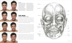
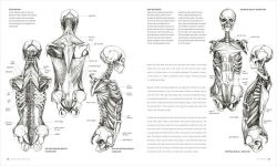
This visually striking guide takes a fresh approach to drawing the human body. A combination of innovative photography and drawings, practical life-drawing lessons, and in-depth explorations of the body's surface and underlying structure are used to reveal and celebrate the human form.

Combining specially-commissioned photographs of models with historical and contemporary works of art and her own dynamic life drawing, Sarah leads us inside the human body to map its skeleton, muscle groups, and body systems. Detailed line drawings superimposed over photographs reveal the links between the body's appearance and its construction. Six drawing classes show how to observe different parts of the body and give expert guidance on how to draw them. Inspirational master classes on famous works, ranging from a Michelangelo study to a Degas painting, show how artists have depicted the human body over the centuries. Each master class includes a photograph of a model holding the same pose as in the painting, to highlight details of anatomy and show how the artist has interpreted them.

Understanding anatomy is the key to drawing the human body successfully. As well as being the perfect reference, Anatomy for the Artist will inspire you to find a model, reach for your pencil, and start drawing.

Product Details

ISBN-13: 9781465494221
Publisher: DK
Publication date: 08/04/2020
Series: Practical Art
Edition description: Reissue
Pages: 256
Sales rank: 260,307
Product dimensions: 9.40(w) x 11.10(h) x 0.90(d)

About the Author

Dr Sarah Simblet is a fine artist, writer, broadcaster and anatomist, whose work explores the relationship between science, history and art. The author of Drawing for the Artist and Botany for the Artist, and co-author of The New Sylva, Sarah is dedicated to sharing and encouraging visual intelligence in others through her drawings, teaching and broadcasting worldwide. She is Director of Studies for Fine Art at Christ Church, Oxford, and Tutor in Anatomy at the Ruskin School of Art. She also teaches short courses in anatomy, botany, and drawing for both the Ruskin School of Art and the Royal Academy of Art, London.

Read an Excerpt

The Forearm and Hand
The dissection and portrayal of the forearm and hand hold a unique and special place in the history of anatomy. Like the brain, they have a significance far beyond the understanding of mechanical function and form. In Christian Europe, the forearm and hand have been seen as God's most profound creation, his tool on earth. The most famous example of this belief is Rembrandt's "Dr. Nicolaes Tulp Demonstrating the Anatomy of the Arm" (1632). The eminent surgeon stands surrounded by intrigued colleagues as he demonstrates the workings of what Aristotle had called "the instrument of instruments."

Hands are supreme instruments of touch. Their sensitivity and delicacy of control make them our primary antennae in out interaction with the word. Their strength and articulation have contributed enormously to our physical environment and to the entire history of artifacts. As organs of highly sophisticated engineering, hands continue to be the subject of profound interests to artists. The contemporary Australian performance artist Stelarc, who builds robotic extensions of his own anatomy, wears a third forearm and hand: a perspex-and-steel device that questions rather than copies the original. In its avoidance of simple mimicry it reflects, like Dr. Tulp, on the meaning and design of this remarkable limb.

The Forearm and Hand Bones
The long bones of the forearm are named the ulna and radius. They lie parallel to each other and are joined at the elbow and wrist. The ulna is placed on the medial or little finger side of the forearm, and the radius is lateral, on the thumb side. They are connected by a fine interosseous membrane, which gives an additional area of attachment to muscles

The ulna is longer, set slightly higher, and behind. Its proximal end is thick and flattened, forming the familiar bony point of the elbow. This point is named the olecranon, and it fits into the olecranon fossa of the humerus, when the limb is held out straight. The anterior surface of the bone forms a tight cup around the trochlea. The olecranon and trochlea meet at a hinged synovial joint. Their movement is restocted to flexion end extension (backward end forward) through one plane. The shaft of the ulna is prismatic (or three sided) throughout most of its length. It gradually tapers, becoming both more slender and cylindrical toward the wrist. The rounded distal end of the ulna (named the head) is excluded from the wrist joint by an articular cartilaginous disk.

The head (or proximal end) of the radius is small, rounded, and flat. It articulates with the capitulum of the humerus above and the radial notch of the ulca to its medial side. It is embraced by a bandlike annular ligament. The shaft of the radius rotates freely across the ulna in its own longitudinal axis. This movement carries the hand from pronation (turning the palm down) to supination (turning the palm upward). Below the head of the radius, the shaft narrows to a slim neck, at the base of which (in front) is the radial tuberosity This supports the insertion of biceps brachii, which flexes and supinates the foreamm. The shaft of the radius is grooved and ridged for the attachment of muscles passing via tendons into the hand. The bone grows wider and flatter toward its distal end, where it articulates with the head of the ulna and the navicular and lunate bones of the wrist. The radius alone carries the skeleton of the hand.

Eight carpal bones shape the wrist. These are small, irregular, and tightly bound by ligaments. Arranged in two curved rows, they are named navicular, lunate, triquetral, pisiform, trapezoid, trapezium, capitate, and hamate. Carpal bones form a short and flexible tunnel, which is bridged and maintained by a strong ligament. The tunnel encloses and protects tendons, blood vessels, and nerves passing beneath. Five metacarpal bones give form and curve to the palm. These articulate at their base with one or more bones of the wrist. At their heads, they support a finger or thumb. The four metacarpals of the palm are tied together by ligaments at their distal heads. The first metacarpal (of the thumb) is independent and rotated through 90 degrees to oppose and press against the palm. There are fourteen phalanges (or finger bones): three in each finger and two in the thumb. Each phalanx (formed of a base, shaft, and head) tapers to give shape and precision to the fingers.

The Forearm and Hand Muscles
The two bones of the forearm are almost constantly in motion throughout our lives, turning back end forth in perpetual service to the hand. Their movement of the wrist and fingers is controlled by more than 30 slender muscles, arranged in layers from the elbow to the palm.

The muscles and accompanying tendons of the forearm and hand fall into three groups. Long extensor muscles shape the posterior of the forearm and pass into the dorsum (or back) of the hand. These supinate the palm and extend the wrist, fingers, and thumb. Flexor muscles cover the anterior of the forearm and pass into the palm. These pronate the palm and flex the wrist, fingers, and thumb. Muscles contained within the palm give mass to the hand, while flexing, extending, adducting, and abducting the fingers and thumb. The long tendons of the forearm divide and insert into numerous bones, giving strength and dexterity to the carps s, metacarpals, and phalanges of the hand.

Muscles and tendons of the forearm are generally named according to their origins and insertions onto bone, relative lengths, and action. Names are therefore often very long. This may seem complicated, but it is actually very helpful in understanding the movement of the limb. For example, flexor carpi ulnaris (far right) flexes the wrist (or carpus) on the side of the ulna. Extensor carpi radialis longus (near right) is the longer of two muscles extending the wrist on the side of the radius. Flexor digti minimi brevis is a short flexor of the littlie finger. Extensor pollicis longus is the long extensor of the thumb, while abductor pollicis longus is the long abductor of the thumb. Palmaris longus is a very long slender muscle tying to the palm.

Of all the muscles of the forearm, palmaris is particularly weak and insignificant. However, it is of special interest because it is often missing, or only present in one forearm. Its absence is sometimes attributed to the refining process of our continuous evolution. Anatomical variations appear throughout the body. Various structures, including muscles and bones, may be either absent a multiplied on one side of the body, perhaps unusually extended, divided, pierced, a united to an adjacent part. When present, palmaris longus can be found in the wrist as a small threadlike tendon, medial to the much thicker cord of flexor carpi radialis.

As muscles contract, they shorten, rise, pull against their tendons, and draw lines of tension between the banes into which they are rooted. Flexed muscles will try to pass in straight lines between their points of origin and insertion, and they are only prevented from doing so by intervening bones, joints, adjacent muscles, and thickened bands of fascia tying their straining fibers into place. More than 20 tendons pass through the wrist. A few of these are visible when taut, and they can seem to press against the skin. They are, however, tied or bound against the bones by thickened fibrous bands named retinacula. Flexor and extensor retinacula enable tendons of the hand to change direction at the wrist, without taking shortcuts across to the forearm.

Tendons inserting within the fingers are similarly bound, to the knuckles. It is important to note that there are no muscles in the fingers -- only tendons on either side of the bones, tied by fibrous bands into lubricated synovial sheaths. The small fleshy bellies of each finger are composed of fatty tissue, carrying blood vessels and nerves, and cushioning flexor tendons against the grip of the hand. Broadly speaking, the dorsum is bony and tendinous, beneath a covering of loose skin. The palm is more muscular and fatty. The muscles of the palm are enclosed beneath a sheet of thickened fascia. Named the palmar aponeurosis, this protects and strengthens the palm. It also ties the skin above to muscles and bones below, holding it firm, and preventing it from slipping when we grasp a surface. The palmar aponeurosis is composed of three parts: a very dense fan-shaped center, flanked by much finer medial and lateral extensions covering the base of the smallest finger and thumb and becoming continuous with the dorsal fascia of the hand. At its apex, the aponeurosis blends with the tendon of palmaris longus, leading in from the wrist. At its base, deep fibres blend with the digital sheaths of flexor tendons and with the transverse metacarpal ligament. Superficial fibres tie to the skin of the fingers and palm, while from its deep surface intermuscular septa pass between muscles of the hand to reach the metacarpal bones beneath.

From Anatomy for the Artist, by Sarah Simblet. © 2001 Sarah Simblet. Used by permission.

Table of Contents

Introduction

The Art of Anatomy
STRUCTURE OF THE HUMAN BODY
Systems
Skeleton
Skeleto-muscular
Integumentary
Respiratory, Digestive, Urinary
Endocrine, Nervous, Lymphatic, Cardio-vascular
BONES & MUSCLES
The Head
Skull Bones
Facial Muscles
Neck Muscles
Ears and Hair
The Spine
The Spine bones
Masterclass: Jean-Auguste Ingres
The Torso
Rib Cage
Muscles
Genitals
Masterclass: Francis Bacon
The Shoulder and Arm
Bones
Muscles
Masterclass: Jacques-Louis David
The Forearm and Hand
Bones
Muscles
Masterclass: Jose de Ribera
The Hip and Thigh
Bones
Muscles
Masterclass: Edward Hopper
The Leg and Foot
Bones
Muscles
Masterclass: Hans Holbein
THE BODY & BALANCE
Poses
The Body in Space
The Model on Paper
Masterclass: Edouard Manet
Movement
Masterclass: Edgar Degas
DRAWING CLASSES
Transparent Bodies
Drawing the Skeleton
The Skeleton in Perspective
Masterclass: Michelangelo
Drawing the Head
Drawing the Rib Cage
Drawing the Pelvis
Drawing the Hands
Masterclass: Raphael
Drawing the Feet
Key Terms
Glossary
Further Reading
Directory
Index
Acknowledgments

From the B&N Reads Blog

Customer Reviews