
eBook
Available on Compatible NOOK devices, the free NOOK App and in My Digital Library.
Related collections and offers
Overview
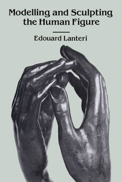
Is there art after modernism? Many of today's art students and professionals are finding the answer — "yes" — lies in the long-neglected field of figurative sculpture, a demanding form of expression that requires extremely rigorous technical training. Most modern schools, however, are simply not equipped to provide the necessary technical background. The republication of this highly valuable text by Edouard Lanteri, renowned teacher, sculptor, and intimate friend of Rodin (Rodin called him "my dear master, my dear friend"), makes it possible for serious students to gain the requisite skills and bridge the gap between artistic concept and figurative realization. Representing at least three thousand years of studio lore, this readily understandable, authoritative guide is a goldmine of technical information, easily comprising a four-year sculpture curriculum unavailable elsewhere.
Beginning with a detailed study of modelling a head from a cast model, Lanteri gives meticulous descriptions of the anatomical features that comprise the head. Next, there are instructions for sculpting a bust from a live model: how to place the model, use the clay, take measurements, set up the all-important framework, put on hair, etc. The author also covers modelling the figure from nature, including such factors as the scale of proportions, posing the model, the chief line, contrasts of line, building up the figure, and more.
Part III covers sculpting in relief (poses, fixing the background, tools, superposition of planes, color, change of light, etc.); drapery (arrangement of folds, principles of radiation, flying drapery, etc.); and medals (proportion, working the mold, inscriptions, etc.). Also discussed are principles of composition, both in relief and in the round. Profusely illustrated with hundreds of photographs, drawings, and diagrams, this work is the kind of comprehensive resource that should be a lifelong studio companion to the figure sculptor. 107 full-page photographic plates, 27 other photographs, 175 drawings and diagrams.

Product Details
| ISBN-13: | 9780486132365 |
|---|---|
| Publisher: | Dover Publications |
| Publication date: | 07/05/2012 |
| Series: | Dover Art Instruction |
| Sold by: | Barnes & Noble |
| Format: | eBook |
| Pages: | 480 |
| File size: | 55 MB |
| Note: | This product may take a few minutes to download. |
Read an Excerpt
Modelling and Sculpting the Human Figure
By Edouard Lanteri
Dover Publications, Inc.
Copyright © 1985 Dover Publications, Inc.All rights reserved.
ISBN: 978-0-486-13236-5
CHAPTER 1
PART I
TOOLS
(i) Provide yourself with two turntables,—one for the work, the other for the model, the height from the ground about 3 feet 4 inches to 3 feet 6 inches. See Fig. 1.
(2) Provide two wooden boards, about 18 inches square or larger, according to the size of your work. To avoid warping through the moist clay, have the boards clamped at the back with two battens nailed or screwed on crossways. See Fig. 2.
(3) A few wooden tools are enough to begin with, the preference to be given to those of simple form; avoid tools which are heavy in your hand, as well as the small and thin ones, unless your work is to be in very small proportions. One tool with strong wire curves at the extremities will be found helpful. For shapes see Fig. 3.
(4) Provide a pair of large iron compasses or callipers, with their legs slightly curved, and about 10 to 12 inches long. These are best ordered from a local blacksmith. Fig. 4.
(5) Have at hand a small sponge to keep your fingers clean during modelling, and some soft old linen to cover up the work after leaving off for the day. Occasionally you will also require a fine syringe or a big brush to splash the clay, when it begins to harden. As we go on, I shall describe whatever tools become necessary; at first no more than the abovementioned are required.
FIRST STUDIES.—THE FEATURES
THE MOUTH
THE best models for the details of the face I consider to be those taken from the mask of Michel-Angelo's "David." They are executed with such precision, so much knowledge of form and anatomy, that in copying them the student is seized with the desire to know the reason for these forms, and he is thus urged on to the study of ANATOMY, so necessary for sculpture.
After having fixed your plaster cast on a board like the one you are going to work on, and having placed it on the turntable, fix both boards at the same angle by means of a wooden support, nailed to the board and turntable, (or by an iron support, screwed on to both), see Fig. 5, so that your work, as well as the plaster model, is in a vertical position. Then drive a nail into your board, at about the level of your eyes. To this nail, by means of some wire (galvanised iron-wire will do, though copper-wire is preferable) fix a small wooden cross ("butterfly" in studio parlance—see Fig. 5), which will help to bear the weight of the clay. Then with your sponge wet the board and immediately rub some clay on the wetted part, so as to form a thickish paste to which your clay will adhere. If you neglect this, your clay will soon get loose from the board and drop down, but this paste and the butterfly will keep it in its place.
Let me just mention by the way, that the more you work the clay through with your hands, the more pliable and consequently the better it will be.
To begin work, lay a certain quantity of clay on your board, pressing it well in with the thumb, so as to give about the general shape and mass of your model, always taking care to keep a little below the actual amount, so that there will be no need to scrape off, but rather to add on, in order to arrive at the true form of the model.
Before beginning each study, you must try to understand what you wish to reproduce, and I shall, therefore, introduce each feature by a sketch of its characteristics and its anatomy.
The mouth is formed by the Upper and Lower Jaw; the upper jaw consists of the two Superior Maxillary Bones; they are two large bones, joined in the middle line and placed almost vertically beneath the Frontal Bone. By means of four processes —the Malar, Alveolar, Palatine and Nasal Processes, which extend in different directions, these bones are joined to the other bones of the face. The Malar processes connect them with the cheekbones. The Alveolar process forms the Superior Dental Arch, which contains the sockets for the Upper Teeth. The Palatine Process passes inwards to form the anterior part of the hard palate, and the Nasal Process extends upwards to the orbits and the Internal Angular Process of the Frontal Bone; it forms the sides of the nose and is connected with the Nasal Bone in front, its outer border forming the deep notch which constitutes the anterior opening of the nose.
The Lower Jaw Bone or Mandible is originally composed of two halves which very early become a single bone, i.e., the Inferior Maxillary. It consists of a solid, horseshoe-shaped body, and upturned, flattened ends, the Rami or Branches. See Photograph of Skull, Figs. 6, 7 and 8.
The posterior border of each ramus expands into an oblong condyle which fits into the Glenoid Cavity of the Temporal bone and forms the Temporo-maxillary Articulation, a very secure double-hinged joint, which allows backward and forward, as well as lateral movement of the lower jaw. The junction of the body of the bone and the Rami forms a rounded angle, which really decides the width and shape of the cheek. Apart from individual varieties, the angle is more acute in the fully developed individual than in the child, or again in an old man, where it is obtuse. See Figs. 9, 10 and 11.
In the same way the Alveolar Processes are not so prominent in a child as in a full-grown person; and in old age, when the teeth fall out, these processes become absorbed and disappear.
There are various processes and ridges on this bone, which serve as attachment to powerful muscles; most important for our purpose is the triangular Mental Eminence, or Chin, which is subcutaneous, and therefore of great service for taking measures from, besides its being a very characteristic feature in the human face. Above it is the Symphysis, a vertical line which marks the union of the two Maxillary bones.
The transverse line which we specially designate by the name of Mouth is surrounded by the lips, which in their turn are surrounded by the Labial group of muscles. These are :—
(a) The Levator of the Upper Lip, which raises it and draws it forwards;
(b) The Levator of the Angle, which raises the outer part of the Upper Lip;
(c) The Zygomaticus Major, which draws the corner backwards and upwards, and is a laughing muscle;
(a) The Zygomaticus Minor, which draws the outer part of the upper lip in an upward, outward, and backward direction;
(e) The Depressor of the Angle, which draws the corner of the mouth downwards and backwards and assists in producing a sad expression;
(f) The Depressor of the Lower Lip, which draws the lower Lip down;
(g) The Levator of the Lower Lip, used in raising it and protruding it; and last—not least
(h) The Orbicularis Oris, which surrounds the mouth and which forms the Lips. On the upper lip it forms a vertical Median Furrow, the ridges of which form the inner borders of two long triangular spaces, the other two sides of these being formed by the free border of the upper lip and the naso-labial line descending from the wings of the nose to the corner of the mouth. Figs. 12 and 13.
The Median Furrow ends in the prominent Median Lobe of the upper lip; at its sides are slight depressions beyond which the lips are carried on by a convex form to the corners. The lower lip has no median projection, but there is a Median depression from which two convex forms start towards the angle of the mouth, where the upper and lower lips join. The skin which covers this red border of the lips is closely adherent. From the fact of there being no cartilaginous framework in the lips, they are very flexible and lend themselves to the most varied expressions.
There is another small surface-muscle, called specially Risorius or Laughing Muscle, which acts together with the Zygomaticus Major, to produce a laughing expression. See Figs. 12 and 13.
* * *
After having well mastered the anatomical characteristics, and having laid on your board a certain quantity of clay, draw on this mass a horizontal line to represent the division of the lips; then with your callipers measure the distance from corner to corner of the mouth, and set this distance off on your horizontal line. Turn your board and the plaster model sideways, and work in profile—with small balls of clay—the outline of the middle with its projections and depressions; do the same from the opposite side-view; then stand in front of the work, and with the work executed from the profile as your guide, fill in the rest,—always keeping slightly below the volume.
You may then at once apply your anatomical knowledge, and lay the superficial muscles on in separate, well-defined strips, rather exaggerating their form than drawing them with indecision. See Photograph of mouth in three stages, Figs. 14, 15 and 16.
And now you ought to proceed by section,&mdsh;that is, by studying your work from below and above,—taking good care that you look at your model and your work from the same point of view, from below and above, so as to compare them and try to obtain the same contours from these points of view. Thus you will correct what you have done so far.
Then look at your work and the model from a three-quarter view, and correct it from that point of view.
Having worked your masses well together from these different points, you proceed to work by light and shade and the numerous half-shades: this I call Colour, and I shall use the expression "colour" in this work entirely in the sense of light and shade. You must be careful to place your work in the same effect of light as the model, and in a strongly projected light from the side, which will give you a greater variety of shades than a light from the front,—in the latter, numerous small depressions pass unobserved, so that the result would be a surface without life and movement.
With the help of this working by colour, and by frequently turning the work and model so as to gain different effects, and different points of view for your drawing, you will at last arrive at the simple form of your model. In short, you must always correct your work by drawing, and must try to obtain simplicity by colour, and expression by your knowledge of the form. Observe that work done in a side-light will always look well when turned to the front; whilst, on the other hand, work done in full light will from a side-view look uneven and undecided in its planes.
Take care not to use the tool too much; it will prevent you from acquiring suppleness of the hand and from developing a fine touch. The human finger, more firm and sure than the wooden tool, will best transmit the intentions of the artist, and express them in varied degrees; the finger is an intelligent, energetic, I might almost say an intellectual, instrument, and you must always use it wherever you can get it in, and only use the wooden tool in places where the finger cannot get in.
SECOND FEATURE.—THE NOSE
The nose is formed partly by bone and partly by cartilage. The two elongated Nasal Bones, joined to the Cranium just below the Glabella, form the bridge of the nose, and are supported at the sides by the ascending processes of the Superior Maxillary; to their anterior border are attached the Lateral Cartilages of the nose. There are five principal cartilages, an upper and a lower Lateral cartilage on each side, and a median one between the two halves of the nose. See Figs. 6 and 7, page 11, and Fig. 12, page 14.
The lower lateral cartilages support the wings of the nose. There is a vast difference in the shape of the nose in races and in individuals, owing to variety of form in the arch, as well as in the cartilage. The muscles but slightly conceal the shape of the bone. The principal muscles from the artist's point of view are:
(i) The Pyramidalis Nasi, which is really only a continuation of the big Frontal muscle;
(k) The Compressor of the Wings, which envelops the sides of the nose below the previous muscle;
(l and m) The very diminutive Anterior and Posterior Dilators of the Nose, at the side of the nose, and hardly visible;
(n) The Depressors of the Wings of the Nose; and
(o) The common Levator of the Upper Lip and Nose. Refer to Figs. 12 and 13, page 14.
When you have well studied the position and characteristics of these bones and muscles, erect your board in the same way (vertically) by the side of your model. To give the better support to your work, make a small butterfly, which ought to be inside the most projecting part of the nose, taking care to make it sufficiently small, so as not to allow the ends to show or stick out of your finished work.
Proceed then in the same way as for the beginning of the previous feature, i.e. moisten your board, make a clay paste on it, and let me say, once for all, that this has always to be done on starting modelling on a background. Having put on a sufficient mass of clay to cover the cross and give the size of your model, you measure with callipers the length of the nose, and at once start to work the profile, first from one side, then from the other, then from below and above, to get the outlines of wings and nostrils sharply indicated; then you take the three-quarter views, and at last the colour, in a side-light, thus modelling and drawing alternately from every point of view, to correct your work wherever you see a mistake, until it is satisfactory. I would particularly warn you not to round off too much the tip of the nose, but well show the planes. See Figs. 17, 18, and 19.
THIRD FEATURE.—THE EAR
For our purpose the outer ear, or Auricle, is all that is needful, whilst the Anatomist proper has to study carefully also the parts of the ear hidden from view.
We have to examine in the Auricle the external and the internal face, and observe that the depressions of one side are the protuberances of the other.
The framework of this feature consists not of bone, but of cartilage, much turned and twisted, which descends right into the Ear-Passage, where it is attached to the bony structure of the skull; the cartilage does not descend into the lobe of the ear, which is made up of areolar and fatty tissue.
The outer curved rim of the ear is called the "Helix"; it is generally more or less folded over at the upper part; within this curve, and parallel to it, is the shorter curve of the Antihelix, a ridge which surrounds the deep Shell or Concha, that leads into the EarPassage or Auditory Meatus. Its upper part branches off into two ridges between which lies a small Fossa, which loses itself under the overlapping part of the Helix.
In front of the Concha, protecting, as it were, the opening into the ear, is the Tragus, presenting a convex form to the outside. Immediately behind its lower part is a deep notch (—the point from which we take our measures for the face—), behind which rises another protuberance, called the Antitragus, and below it extends the Lobule or Lobe of the ear. The muscles which lie on the Tragus and Helix are very insignificant, even the three Auricular muscles, which fix the ear to the skull, are so small and thin, that they hardly influence the surface-form. The ear is covered with a fine and smooth skin, which closely follows all the depressions and projections of the cartilaginous framework. (Fig. 20.)
The process of working will be the same as indicated for the previous studies; only let me repeat that the principle of drawing and working by colour-effect is the only principle I shall insist upon for all our studies, whether they be from the cast or from nature. That is why a modelling-student must above all be a good draughtsman, for drawing will give him not only precision in the form, but it is also the only means of making his work graceful and telling,—a quality of work which can never be obtained by him who cannot draw, and whose work will always remain heavy and commonplace. Therefore when you have understood the why and wherefore of each form, and know it by heart, drawing will remain the most important part of your work. See Figs. 21, 22, and 23.
(Continues...)
Excerpted from Modelling and Sculpting the Human Figure by Edouard Lanteri. Copyright © 1985 Dover Publications, Inc.. Excerpted by permission of Dover Publications, Inc..
All rights reserved. No part of this excerpt may be reproduced or reprinted without permission in writing from the publisher.
Excerpts are provided by Dial-A-Book Inc. solely for the personal use of visitors to this web site.
Table of Contents
INTRODUCTIONPART I
TOOLS
THE FEATURES FROM THE CAST
(a) The Mouth
(b) The Nose
(c) The Ear
(d) The Eye
THE HEAD FROM THE CAST
(I) The Skull
(2) The Muscles
PART II
THE BUST FROM LIFE
MEASUREMENTS
DIAGRAM OF CONSTRUCTION OF FEATURES
(a) Mouth
(b) Nose
(c) Ear
(d) Eye
TREATMENT OF HAIR
MOUSTACHE AND BEARD
DIAGRAM OF FACE
PART III
FIGURE FROM NATURE
FRAMEWORK
SCALE OF PROPORTIONS
POSING THE MODEL
CHIEF LINE
CONTRASTS OF LINES AND OF PROJECTION OF SURFACES
BUILDING UP OF FIGURE
MEASUREMENTS AND OSTEOLOGICAL CONSTRUCTION OF FIGURE
INFLUENCE OF OSTEOLOGY ON SUPERFICIAL FORMS
RADIATION OF LINES
EXAMPLE OF THE ILISSUS
SPACES OF REST BETWEEN MASSES
EXPRESSION IN MODELLING
CONTINUATION OF LINES
COMPARATIVE PROPORTIONS
MUSCLES OF THE FIGURE
PHOTOGRAPHS FROM CLAY MODEL AND DIAGRAMS
THE SKELETON IN DIFFERENT VIEWS


